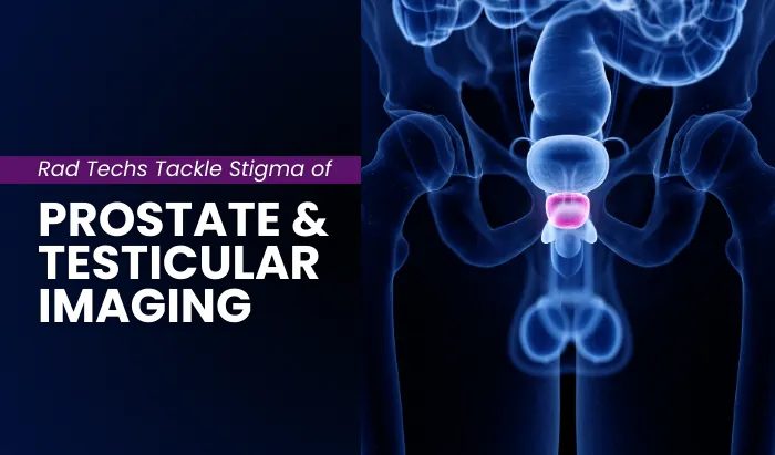
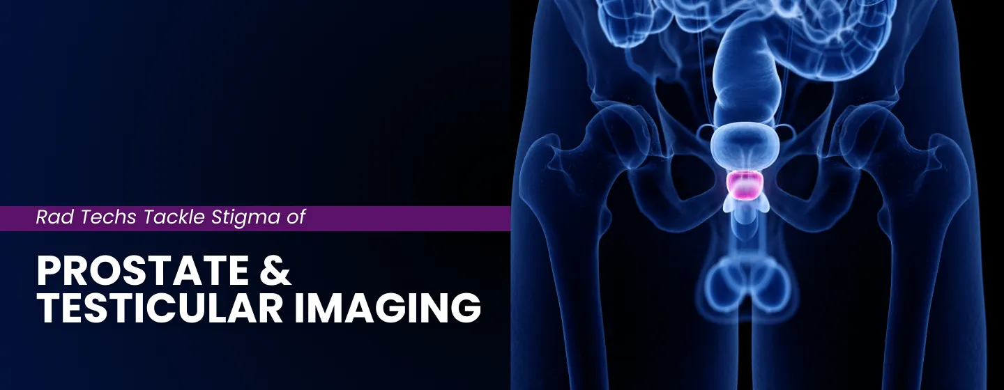
Rad Techs Tackle Stigma of Prostate & Testicular Imaging

- Introduction
- What men commonly feel about prostate and testicular imaging, and why?
- Why delaying imaging procedures is dangerous (the reality of early detection)
- A review of prostate and testicular imaging techniques and patient experiences
- Prostate MRI
- Transrectal ultrasound (TRUS)
- Scrotal ultrasound
- How to make men feel more comfortable during these procedures?
- Before the appointment (patient preparation & scheduling)
- Upon arrival and pre-exam (set the scene, education, privacy)
- During the procedure (narration, permission checkpoints, comfort)
- Professional standards and ethics
- Conclusion
- References
Introduction
Men often delay or avoid testicular, prostate, and colon imaging because of embarrassment, discomfort, and the stigma around these exams. Prostate MRI, transrectal ultrasound (TRUS), and scrotal ultrasound are necessary but intimidating procedures for male patients. As the face of imaging, radiologic technologists can transform these tense encounters into trustworthy experiences through small and personalized approaches that protect patient privacy, normalize the examination process, and create a sense of comfort and ease, all while maintaining image quality.
In 2025, the American Cancer Society (ACS) estimates approximately 313,780 new prostate cancer cases and almost 35,770 deaths in the United States. Testicular cancer is far less common, about 9,720 new cases, but it is more frequently diagnosed because it is often present as a palpable mass that prompts evaluation. Rectal cancer alone accounts for about 46,950 new cases in 2025 (within the broader colorectal category).

What men commonly feel about prostate and testicular imaging, and why?
Many men arrive at their procedures anxious and guarded, already bracing for how the appointment will go. As radiologic technologists, recognizing these emotions upfront helps us choose language, pacing, and privacy measures that defuse tension. Here are the feelings we see most often during prostate, testicular, and rectal imaging and the reasons behind them:
- Embarrassment/fear of judgment. Many men feel uneasy discussing intimate symptoms and may withhold details once in the room, driven by stigma and worry about being judged.
- Discomfort with intimate exams. Being examined in the scrotal or rectal region can feel exposing and distressing, especially in unfamiliar settings.
- Uncertainty about the process. Not knowing what will happen, how long it takes, or what sensations to expect increases anticipatory anxiety.
- Privacy worries. Concerns about exposure during the exam and about who can view images or access information in the record.
- Fear of a cancer diagnosis. Anxiety about “bad news” contributes to postponing care, pushing evaluation later in the disease course.
- Pain/comfort concerns include worries about discomfort during scrotal ultrasound, TRUS, or lying still for MRI, which may lead to cancellations or no-shows.
Why delaying is dangerous (the reality of early detection)
Delaying imaging appointments, ignoring early prostate and testicular screening recommendations, and avoiding routine checkups can be dangerous. Here’s why early detection is a lifesaver:
Testicular torsion (time = testis)
Salvage is ~97–100% within the first 6 hours; viability falls to ~42% by 19–24 hours and approaches 0% beyond a day. Late presentation risks testicular loss and downstream fertility impact.
Prostate cancer (stage drives survival)
Five-year relative survival is >99% when the disease is localized or is found to be regional, but drops to ~37% when distant metastases are present, one reason why timely work-ups of urinary symptoms and follow-through on imaging are crucial.
Avoidance is common (and fixable)
In a national survey, 72% of men said they’d rather do household chores than see a doctor, and 20% admitted they weren’t fully candid once there, behaviors that fuel delay right at the imaging doorstep. Normalizing sensitive exams helps convert avoidance into attendance.
A review of prostate and testicular imaging techniques and patient experiences
Patient experiences in imaging procedures vary between modalities and techniques, but a few stand out as more uncomfortable for men than others. Below is a brief overview of such exams and the reasons why patients may feel uneasy or embarrassed.
Prostate and rectal MRI
Magnetic resonance imaging is a cross-sectional imaging of the internal organs in the body using a strong magnet and radiofrequency pulses to produce different sequences and imaging views. Patients can experience discomfort due to several factors during MR imaging of the prostate, genitals, or rectum, and here is why:
- Enclosed bore & noise: Patients lie in a narrow MRI bore with loud gradient noises for 25–40 minutes, which can trigger claustrophobia or anxiety.

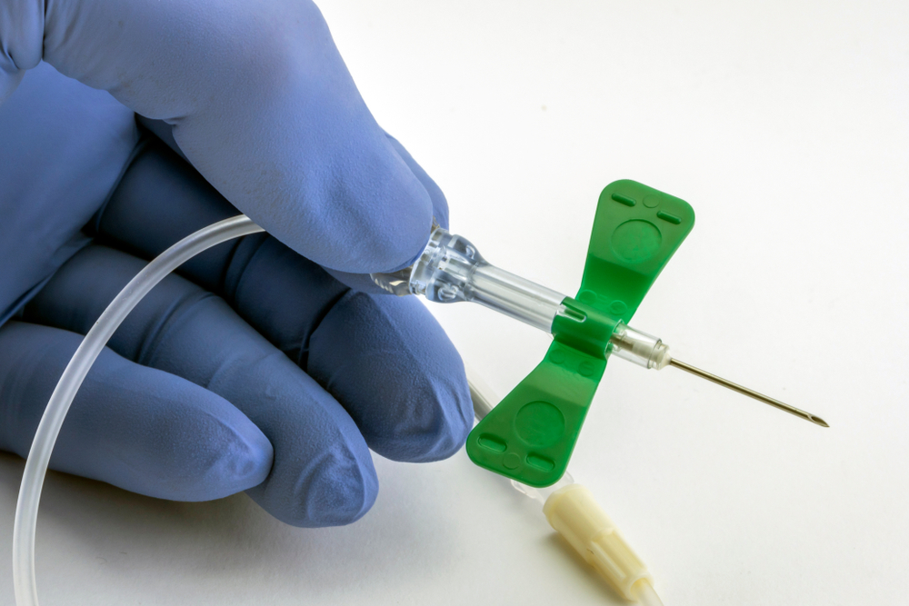
- IV placement for contrast: If gadolinium-based contrast is ordered, an IV needle is required, and it is often a source of anticipatory fear or discomfort.
- Possible endorectal coil: Some centers use an endorectal (rectal) coil, which is a small, flexible coil placed in the rectum, to improve signal and detail. Others rely solely on an external pelvic/phased-array coil. The possibility of a rectal coil can induce embarrassment or apprehension.

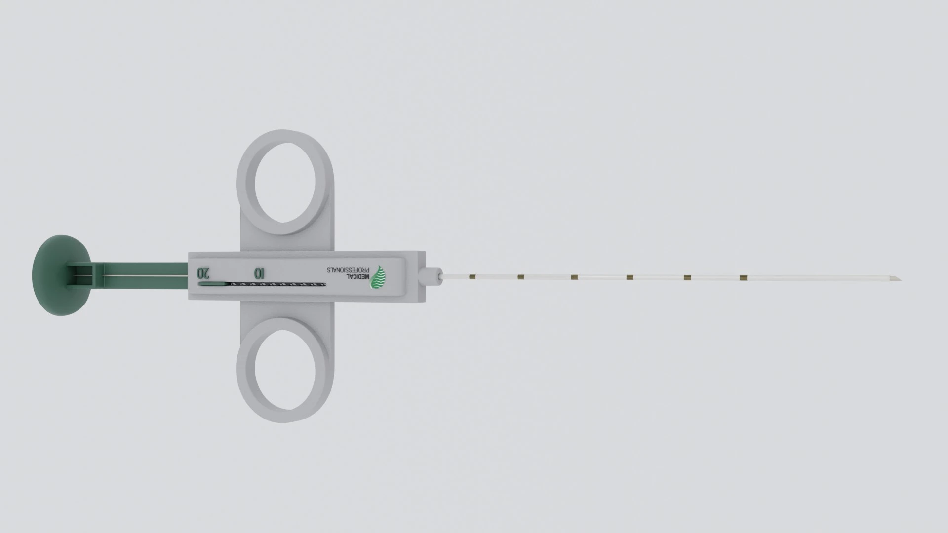
Transrectal ultrasound (TRUS)
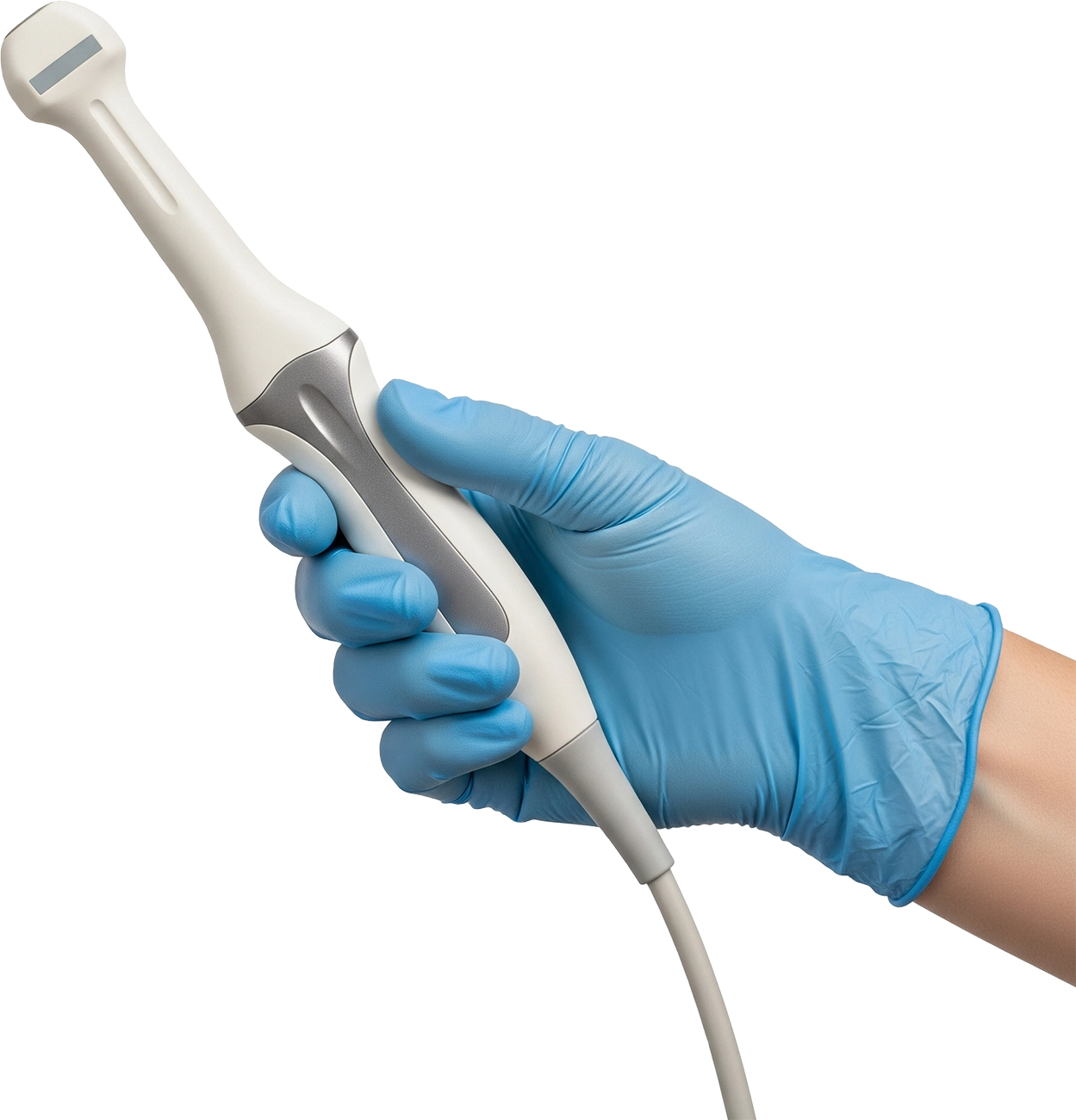
Transrectal ultrasound (TRUS) uses a small rectal probe to visualize the prostate and peri-prostatic tissues (it may also be used for guided biopsies). Patients can find TRUS distressing for several reasons, including:
- Rectal probe insertion & pressure: The sensation is typically a pressure or fullness rather than sharp pain. But anticipating a rectal insertion can heighten embarrassment or anxiety.
- Exposure & positioning: Targeted exposure of the buttocks or perineum in left lateral decubitus (or lithotomy per urology) can feel vulnerable, especially if multiple staff members are present.
- Lubricant/lidocaine jelly: The cool gel or lidocaine jelly used for comfort can feel unfamiliar and increase self-consciousness.
- Biopsy: In cases of a biopsy, patients may worry about needles, pain, bleeding, or hearing the biopsy device “click,” which can amplify apprehension.
Scrotal ultrasound (with Doppler)
Scrotal ultrasound is the first-line study for scrotal pain, swelling, palpable mass, or suspected torsion; grayscale imaging is combined with color/spectral Doppler to assess perfusion. Patients may feel uncomfortable for several reasons:
- Genital exposure & draping: Even with a towel “hammock” and selective draping, targeted exposure of the scrotum can feel vulnerable or embarrassing.
- Probe pressure on a tender area: Warm gel and transducer contact over a painful testis (e.g., torsion/epididymitis) can heighten discomfort despite gentle technique.
- Doppler sounds & stillness: Brief Doppler audio and the need to hold still during discomfort can increase anxiety.
- Fertility/sexual-function worries: When a palpable mass or severe pain prompted the exam, patients may fear implications for fertility or sexual function, adding emotional stress during imaging.
How to make men feel more comfortable during these procedures?
Before the exam starts, the radiologic technologist’s tone and the room’s setup shape the patient’s mindset, driving cooperation and ultimately, image quality. Comfort, privacy, plain-language explanations, validation of concerns, and brief permission check-ins lower anxiety and improve communication. Sometimes, the smallest details (often unnoticed when everything goes right) make the biggest difference.
- Before the appointment (patient preparation & scheduling)
- When booking, a clear scheduling script: “This is a sensitive study. Your privacy is a priority. You’re welcome to bring a chaperone or support person if you’d like. You’ll change in a private space, and only the area being imaged will be exposed.”
- Confirm site-specific prep (arrive early for forms, bring current meds list, empty bladder vs not, bowel prep if your protocol uses one for prostate MRI/TRUS).
- Offer support through an open line for questions or concerns with a radiologic technologist or a sonographer, before, during, and after the appointment.
- Reminder text/email (patient preparation instructions): Reiterate time, location, clothing guidance (easy on/off), specific preparation if needed, and chaperone availability. Keep the instructions understandable and easy.
- Pre-visit materials (brochure/video/leaflet): Send a short, simple overview of the exam (prep, step-by-step what to expect, simple visuals). This reduces “unknowns” and first-visit anxiety; tag this along with the reminder message (captions and multilingual if available).
- Upon arrival and pre-exam (set the scene, education, and privacy)
- Set the scene: Door closed, curtain drawn, gown folded on the chair, drapes visible. Briefly say who will be in the room, how long any exposure lasts, and where the images go. Use phrases like: “Only what we need to see will be uncovered, and only for as long as needed.”
- Patient education (brief reassurance): What the exam does, sensations (warm gel, gentle pressure, machine sounds), duration, and result pickup. Use simple explanatory phrases like: “You’ll hear a short Doppler sound next, that’s normal.”
- Use a model/diagram + medically accurate language: A quick point to a small anatomy model or a poster diagram to show the probe position on the region of interest. Use neutral clinical terms: testis, scrotum, perineum, prostate, and no slang or jokes; example: “Here’s the area we’re imaging and how the probe will sit.”
- During the procedure (narration, permission checkpoints, comfort)
- Explain as you go: Quiet, matter-of-fact narration: “Starting on the right… Now the left… brief Doppler next.” Warm gel when possible and reduce pressure if pain flares. Use a phrase like: “If anything hurts, tell me, and we can pause.”
- Permission checkpoints: Short pauses before a new step or any change in exposure or position: “Okay to proceed?” These checkpoints take a few extra seconds but reduce startle and movement, and give patients a sense of control over their own procedures.
- Draping discipline: Expose only the region of interest and recover immediately after each step. Minimize the duration of exposure as much as possible. Also, minimize room traffic while keeping conversations clinical and focused.
Set expectations early so the visit feels predictable upon arrival.

Prepare the imaging room so that “privacy” is immediately apparent upon entry.
Predictability lowers anxiety; brief pauses give control.
Professional standards and ethics
Use the following standards-driven checklist to hard-wire dignity, privacy, and clarity into every exam. Make dignity and patients’ comfort part of quality, not an add-on. Here are some final, standards-based recommendations for everyday practice:
- Anchor to standards: Focus on privacy, allow chaperones, include consent pauses, and provide clear communication as routine professional practice aligned with:
- ARRT® ethics
- ASRT® Practice Standards
- AIUM Practice Parameter for the performance of ultrasound evaluations of the prostate (and surrounding structures)
- NRG Oncology / MRI protocol
- ACR® parameters
- American College of Radiology ACR Appropriateness Criteria® Pretreatment Detection, Surveillance, and Staging of Prostate Cancer
- Document the key elements: Such as consent forms, patient history, patient’s identifications, chaperone status/name, privacy steps/draping maintained, consent checkpoints, patient preferences honored, and any comfort accommodations.
Quick tip: Standardize this with a short checklist within your protocol so it’s fast, consistent, and auditable.
Conclusion
Radiologic technologists play a central role in making examinations both tolerable and easy, through comforting patients, offering clear explanations, and shaping the experience before, during, and after the study. By destigmatizing sensitive exams and deliberately addressing the feelings we commonly see, like embarrassment, fear of judgment, uncertainty, privacy worries, and pain concerns, we turn avoidance into cooperation. When compassion and protocol work together, we help men move from hesitation to answers and deliver care that is respectful, consistent, and ready for the next patient who needs us.
References (U.S. sources)
- Cancer Facts & Figures 2025 (PDF). (Tables for new cases/deaths; combined colon+rectum mortality.)
- USPSTF (2018, current). Prostate Cancer: Screening Recommendation (55–69 shared decision-making; recommend against routine screening ≥70).
- AUA/SUO Guideline. Early Detection of Prostate Cancer
- ACR Appropriateness Criteria®: Acute Onset of Scrotal Pain—Without Trauma (US with Doppler “usually appropriate”).
- AIUM/ACR/SRU/SPR. Practice Parameter for the Performance of Scrotal Ultrasound (specs, QC, infection prevention).
- SEER (NCI). Cancer Stat Facts: Testicular Cancer (U.S. incidence; age distribution; median age 33).
- CDC (2024 brief). Testicular Cancer Incidence by Tumor Type and Age (three-quarters of cases occur ages of 20–44).
- Cleveland Clinic survey. Men’s Health: Avoidance and honesty patterns in care-seeking. (Survey summary showing avoidance/underreporting.)
- AARP summary of the same survey. Many men would rather do chores than see a doctor; some are not fully candid.
- ARRT® ethics
- ASRT® Practice Standards
- AIUM Practice Parameter for the performance of ultrasound evaluations of the prostate (and surrounding structures)
- NRG Oncology / MRI protocol
- ACR® parameters
- American College of Radiology ACR Appropriateness Criteria® Pretreatment Detection, Surveillance, and Staging of Prostate Cancer
- https://seer.cancer.gov/statfacts/html/colorect.html#:~:text=at a Glance-,At a Glance,65.4%25 2015–2021
Disclaimer: The information provided on this website is intended to provide useful information to radiologic technologists. This information should not replace information provided by state, federal, or professional regulatory and authoritative bodies in the radiological technology industry. While Medical Professionals strives to always provide up-to-date and accurate information, laws, regulations, statutes, rules, and requirements may vary from one state to another and may change. Use of this information is entirely voluntary, and users should always refer to official regulatory bodies before acting on information. Users assume the entire risk as to the results of using the information provided, and in no event shall Medical Professionals be held liable for any direct, consequential, incidental or indirect damages suffered in the course of using the information provided. Medical Professionals hereby disclaims any responsibility for the consequences of any action(s) taken by any user as a result of using the information provided. Users hereby agree not to take action against, or seek to hold, or hold liable, Medical Professionals for the user’s use of the information provided.