
Extravasation of Contrast: The Scope, Magnitude & Gravity of the Problem, and the STEM solution.
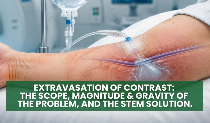
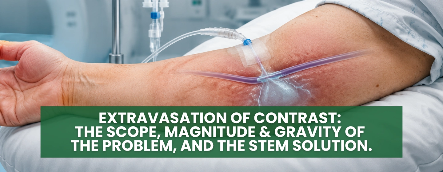






Table of Contents
- Introduction – The Scope, Magnitude, and Gravity of the Extravasation Problem
- Risk Factors: How does extravasation occur?
- Prevention Techniques: Can any change in the execution of this venipuncture and infusion process reduce, or even eliminate, extravasation?
- Early Detection: What are the visible and invisible indicators that extravasation of contrast is occurring?
- Appropriate Management of Extravasation of Contrast – if it occurs.
Introduction
In this article, you are going to read information about extravasation that you have probably never read or heard about before, #1 because extravasation has never been viewed from the Scope, Magnitude, and Gravity perspective before, and #2 because a STEM analysis has never been done before on the venipuncture & infusion of contrast and the complications, compromises, injuries, and failures that arise from this procedure, including extravasation.
Extravasation has been viewed as ‘a minor inconvenience, a problem that can’t be fixed or accepted as a ‘given’, just a ‘fact of life’ in the medical world, and the patient recovers anyway’. But the reality is, this problem is much bigger and more serious than that because extravasation is a serious Side Effect – with consequences.
The Scope
This ‘leakage’ goes by many names, within three different disciplines; over many venipuncture procedures.
This leakage is also called: extravasation, transudation, exudation, and infiltration. And the three disciplines are Nursing, Radiology, & Laboratory Medicine. And it includes these procedures: IVs, Blood Draws, Blood Donations, Plasma Donations, PICC line insertions, Blood Transfusions, Factor VIII Injections, and the Injections of Contrast. (AND no one is looking at this as a global medical problem.)
In fact, any venipuncture that starts with a tourniquet ‘begins and ends’ with an extravasation – it’s a Starling’s Equilibrium fact – to be described later in the article. In Radiology, it’s an extravasation of contrast; in Nursing, it’s an Infiltration; and in Laboratory Medicine it is a ‘pre-analytical phase’ hemoconcentration of the blood specimen – due to extravasation.
The Magnitude
99-100% of all injections of contrast result in some degree of extravasation – due to the use of the TOURNIQUET – which is used 99-100% of the time.
No other invasive, surgical procedure in the practice of Medicine has this kind of Side Effect/Incidence rate. Indeed, if any other medical procedure had a Side Effect rate this high, it would be halted immediately.
Included in the Magnitude are all the other complications, compromises, injuries, and failures (CCIFs) associated with vein access (venipuncture) and infusion. Extravasation is only ONE of the many CCIFs that result from current (19th-20th century) venipuncture methods.
The Gravity
If contrast is extravasating out of the venous system, into the subcutaneous tissue, then not ALL of the contrast ends up in the target organ that is to be visualized and evaluated. Therefore, the question arises – ‘Is there poor perfusion because the organ is ‘sick’ or poor perfusion because some part/amount of the contrast is sitting out in the subcutaneous tissue, instead?’ Now we must question the accuracy of the Radiologic Impression.
Obviously, wrong results lead to a wrong Diagnosis, which leads to a wrong Treatment, which then leads to an adverse Medical Outcome. That’s pretty grave.
Not to mention the potential noxious injury that contrast can cause to the tissue. And how long will it take for the extravasation to be absorbed and removed from the SubQ tissue?
And a final compounding factor contributing to the gravity is that venipuncture is NOT regarded as, taught as, or executed as an invasive surgical procedure – and it is. The root foundation for all venipuncture is the word ‘Phlebotomy’ (-otomy – means surgery, to cut with a scalpel, or, in this case, to penetrate with a sharp instrument, the needle.) Just imagine if this skill were regarded as a surgery; how different would the performance be?
The Scope, Magnitude, and Gravity of this one SIDE EFFECT – extravasation – must be addressed and corrected.
Found this article useful? You may also like these courses.
Explore our hands-on MRI, X-ray and ultrasound courses designed to elevate your clinical expertise and confidence.


Risk Factors – How does extravasation occur?
There is a huge misconception about this aspect of the venipuncture procedure, as well. There are several factors to be considered – from a Science, Technology, Engineering, and Math perspective. Let’s begin.
Most medical personnel think that the needle must be outside of the vein and the contrast is being infused directly into the SubQ, or that the leakage is occurring at the needle insertion site – for some reason.
Here are the real risk factors for causing an extravasation:
- Tourniquet
- Gravity-dependent position of the site
- Vein selection – whether a new site or the use of an already established IV site
- Infusion rate
- Needle Angle of Entry
Let’s start with the Tourniquet. Unbeknownst to most medical personnel, the tourniquet does not ‘dilate’ a vein; the tourniquet forces an overdistention of the vein. This overdistention of the vein thins the walls of the vein, venule, and venous membrane part of the capillary bed (think of an overdistended long skinny water balloon – watch the wall thin as it distends.) Under that increased vascular pressure, from the overfilling with blood, these thinned walls begin to leak – extravasation begins.
This ‘anatomical and physiological’ fact is built upon Starling’s Principle – Starling’s Equilibrium.
In 1896, Ernest Starling described Starling’s Equilibrium. Dr. Starling tells us that there is a pressure balance between the intravascular and the extravascular. And, when there is an imbalance, there is an influx or an efflux of fluid to the system or from the system, respectively.
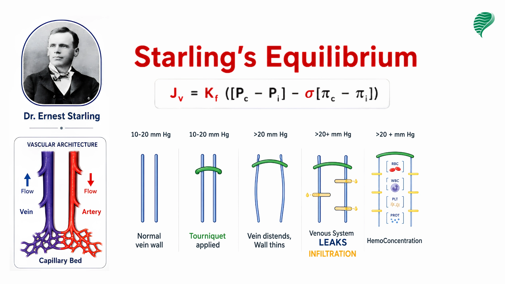
Long story short, when the tourniquet is applied, that external force/pressure forces over distention of the vein, venule, and venous capillary bed, where varying degrees of extravasation occur (most profusely at the venous capillary bed, because of the very thin, porous capillary membrane) – all dependent upon how tight the tourniquet is and for the length of time it is applied; and on the thickness/thinness of the vein wall and the integrity of that vein wall (varicose). 1
The Tourniquet was ‘designed’ to cut off the flow of arterial blood – it never was designed to ‘dilate a vein’.
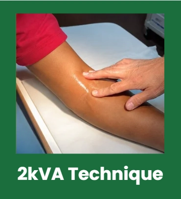
There is a new ‘gentle touch’/palpation method for locating, dilating and grading veins. Gentle Touch causes the innervated smooth muscle media to relax, allowing for vein filling and a natural distention – to the size nature intended for the vein to enlarge to.
If Starling knew, in 1896, that medical people were going to be inserting needles into veins, I am certain he would have told them, ‘DO NOT put a tourniquet on that venous system – it will cause extravasation’.
If in doubt of the veracity of this information, recall the medical profession already uses Starling’s Equilibrium when the physician created that rule about the Mastectomy Axillary Lymphadenectomy patient – ‘no BP, no IV, no blood draws in the affected arm’, because of the resultant leakage (lymphedema). The absence of lymph nodes did not cause the problem. The increased venous pressure caused the leakage.2. What increased the venous pressure? The Tourniquet and the BP cuff. So, P.S., the Rule should have read: No Tourniquet, No BP cuff. It is the TOOL that is causing the problem for that venous system.
Well, that same leakage that occurs in the MALA patient is occurring in EVERY patient – including the injection of contrast patient.
Hmm, that begs the question, “How do you dilate a vein without a tourniquet? Do we need to dilate a vein? Do we need to dilate a vein to locate a vein?” There are answers to these questions, as well. Another article.
Now let’s talk about the Gravity-dependent position of the infusion site. Most IV sites are placed in the hand, some in the wrist, and some antecubital. But ALL rest below the level of the heart (figuratively, and in some cases, literally) – while contrast is being infused.
Where does Gravity take EVERYTHING (including blood and contrast)? DOWN. How is the venous blood and infusing contrast going to return to the heart (and then on to the target organ) if GRAVITY is keeping everything down?
Recall, venous blood only returns to the heart with MOVEMENT – the contracting of skeletal muscles squeezes the vein and ‘milks the blood along’ back to the heart, with the help of ‘valves’ (that hold the blood in place, if you stop moving, till the muscle contracts again).
That means the patient must CONSTANTLY ‘milk the hand’ and move the blood and contrast along. That’s not a reasonable request or expectation. So, what can we do?
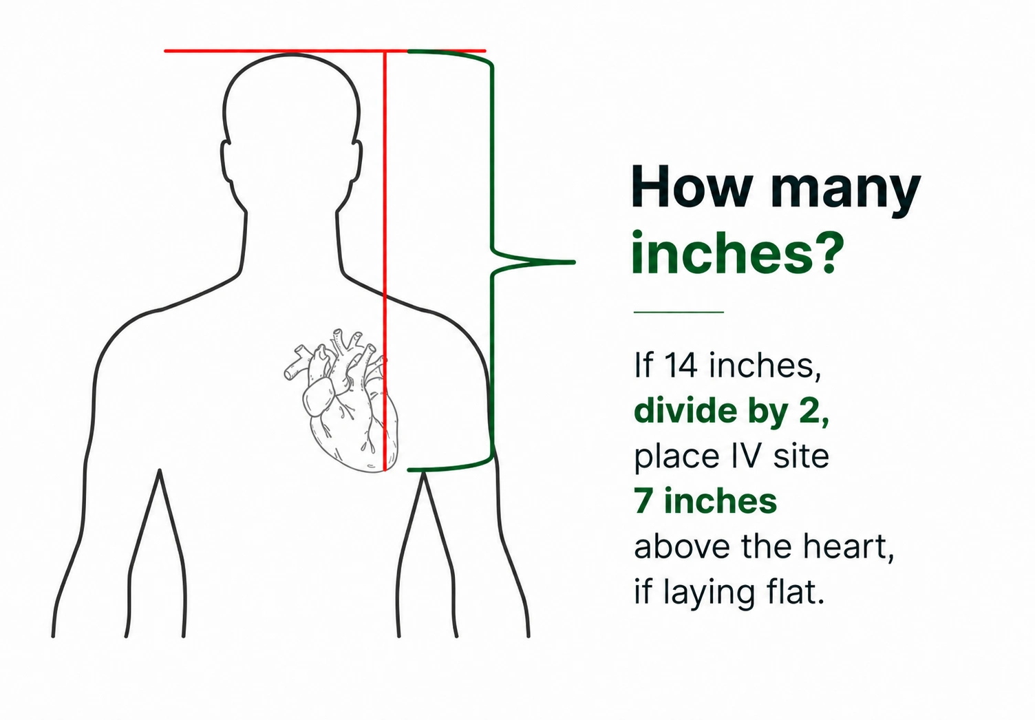
Use Gravity in your favor. Elevate the IV site to between the level of the patient’s heart and the level of their head. You can determine that position by measuring the distance between the patient’s left ventricular apex and the top of their head. The IV site must be elevated at a minimum of at least 4 inches above the level of the heart (whether in the prone position or sitting).
Now GRAVITY will return the venous blood and the contrast to the heart.
If you do NOT help the blood and contrast along, the blood and infusing contrast fluids will just ‘sit’ in that segment of vein – continuing to overfill that segment – which, again, thins the walls of the vein, venule, and venous capillary membrane – resulting in leakage – extravasation.
The next contributing factor to the likelihood of extravasation occurring is Poor vein selection. Not all veins are created equal. Vein wall thickness varies throughout the body. The inferior and superior vena cava have the thickest walls and are closest to the heart; the hand veins have the thinnest walls and are most distal to the heart. The wrist veins and then antecubital veins get thicker, respectively, as they get closer to the heart.
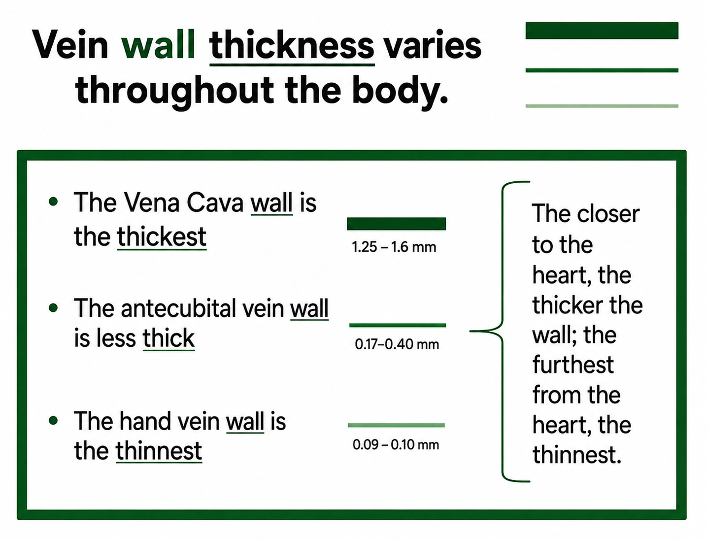
Recall – thin-walled veins leak, along with their venules and venous capillary beds.
Now, imagine putting a tourniquet on an already thin hand vein – and distend it. How often does an IV infiltration/extravasation of contrast occur in the hand?
And you must consider the integrity of the vein that you have selected. You heard of a varicose vein. Those vein walls were overdistended at some point in time and were permanently injured – permanently overstretched – and are now permanently ‘thinner’. How fragile would that compromised vein be if you add the pressure of the tourniquet over distention?
And if you are using an already established Nursing IV site – you must consider all the CCIFs that are probably already associated with that.
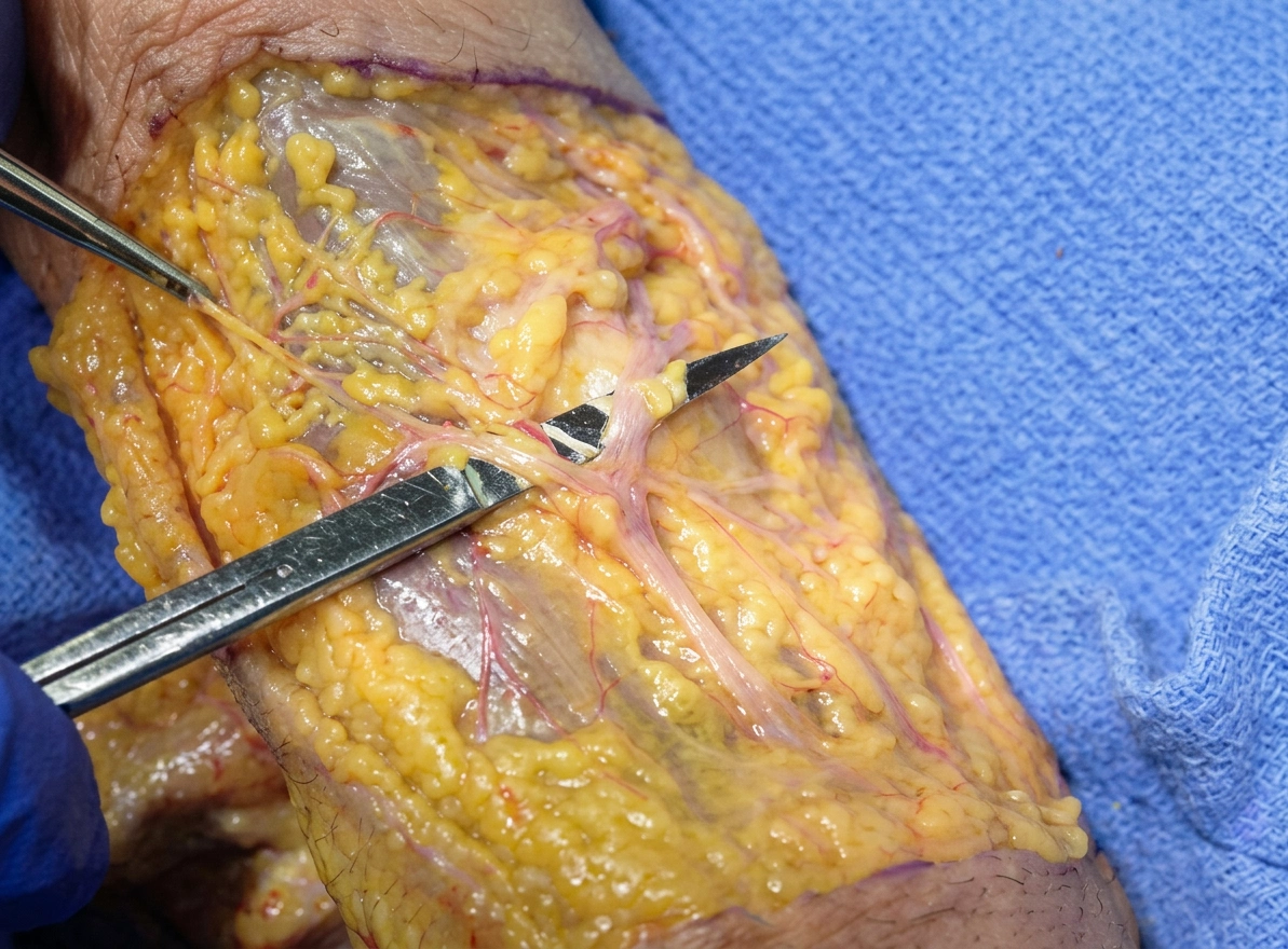
Select a ‘healthy vein’. There is a new ‘Grade the Vein’ technique to determine wall thickness, size, direction, depth, and vein wall integrity. [Adding a gross Anatomy dissection to the training would be helpful.]
How does the rate of infusion factor into all of this? Is the contrast injector a good idea? Is it even needed? How fast, or slow, should that contrast be infused?
Two things to think about here: 1) What amount (mls) of contrast can be injected into that segment of vein before ‘distention’ occurs – and the leakage begins? 2) How fast is the blood flowing through that vein?
[Think of automobiles, rush hour, bumper to bumper, traveling at 55mph – but oncoming traffic (from an ‘on’ ramp) tries to join in at a speed of 75mph – what will happen? There will be a collision, car will bunch up, bulge, and spill out onto the shoulder.]
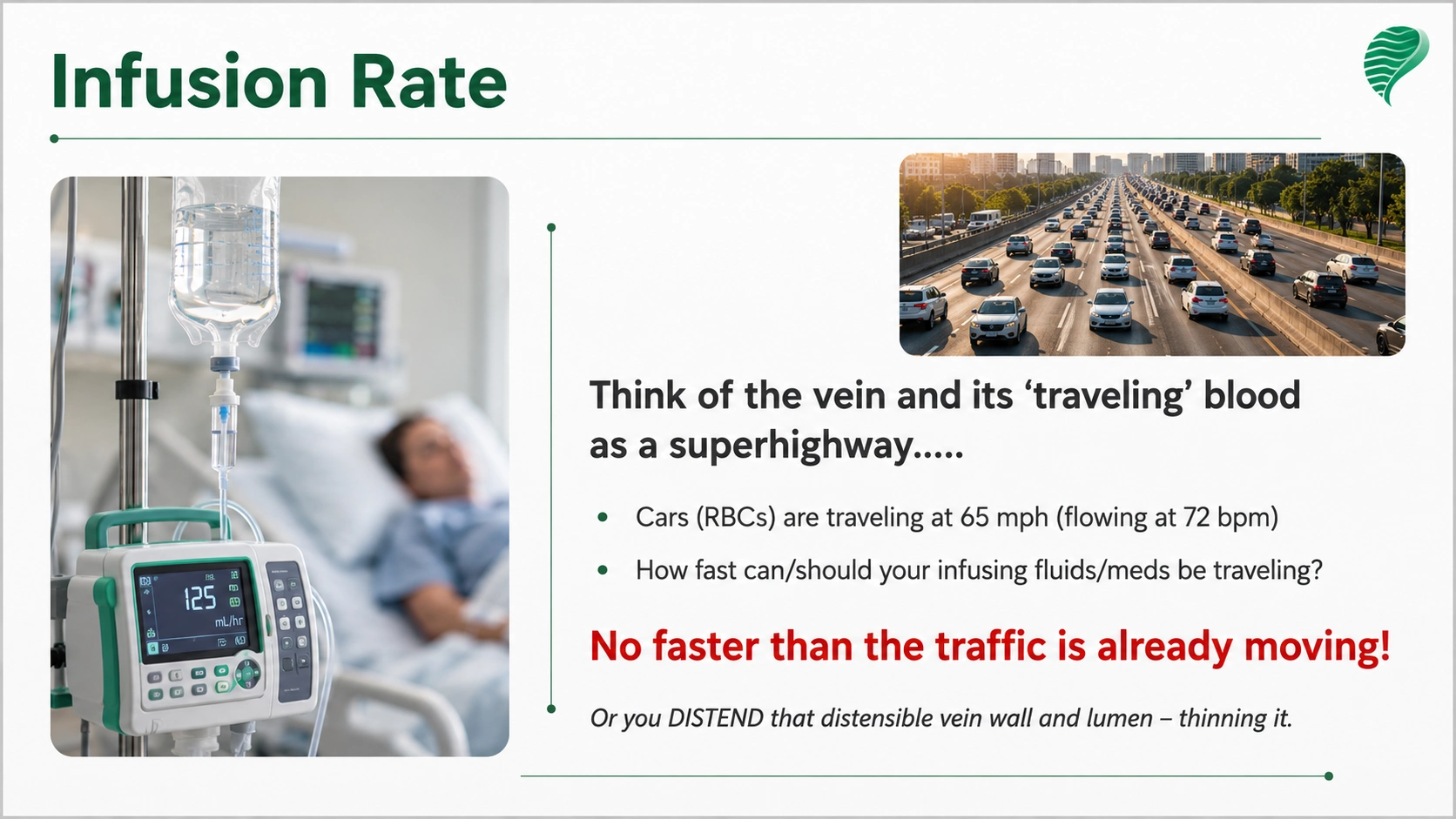
If the contrast liquid is infused faster than the blood is traveling (patient’s heart rate), then you will cause an overfilling and an overdistention of the vein (venule, and venous capillary bed) at that site, causing leakage/extravasation.
Injectors have advanced the safety and accuracy 4 – but focus on the speed at which the contrast is injected – just add the patient’s blood ‘travel’ consideration to the use of the injector speed and power behind the actual infusion.
One last factor to consider is the needle angle of entry through the vein wall and into the lumen of that vein. The 15o-30o degree typical angle of entry cuts a lot of vein wall – weakening that anatomical structure. And if unnecessary external pressure (from a tourniquet) and internal pressure (from a rapid infusion and gravity ‘pooling’) are applied to that weakened wall, the cut extends, and the leakage begins. Compare the two entries above – which causes less injury?
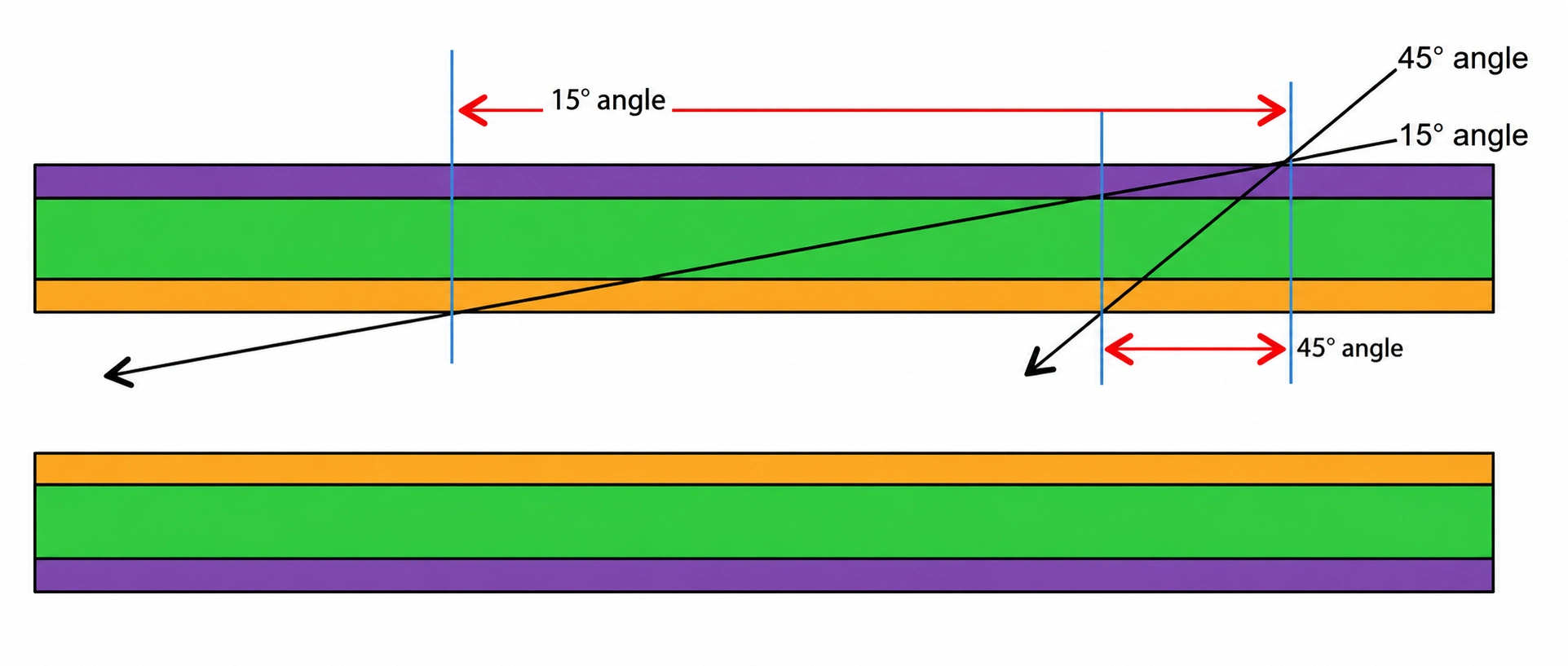
And instead of gauging flow rate based upon the gauge of the needle, the selected vein determines the gauge of the needle to be used – adjust external flow rates to the size of the needle AND to the patient’s ‘flow rate’.
If you have gravity working in your favor, without a tourniquet to locate a vein, with a healthy vein wall, right-size needle for that vein lumen, correct needle angle of entry and appropriate quantity/speed of contrast in congruence with the patient’s physiology – there will be no extravasation.
How then can that extravasation be prevented? Prevention Techniques:
- Select a healthy vein – grade for wall thickness and integrity.
- Do not use a Tourniquet. If venous ‘support’ is needed, there is a new tool on the horizon to support the venous system (not distort it, as the tourniquet does).
- Infuse slowly – with the speed of the blood in mind (heart rate and the speed at which the blood is traveling in the vein).
- Insert the needle at a 45 degree angle of entry – then place the catheter in an indwelling (flat) position.
Early Detection of Extravasation of Contrast
Invisible Signs: First to appear. Patient c/o burning sensation. (If you are using an already established Nursing IV site that is already infiltrating, this invisible sign may not be easily recognized by the patient.)
Visible sign: Second to appear – swelling. But this sign is late in the game, usually after the patient has left radiology.
Appropriate Management – if extravasation occurs.
Nursing will need to alternate cold/warm packs to the site. Cold to reduce swelling from tissue injury. And Heat to increase circulation to start the reabsorption of contrast and fluids back into the vascular system, because macrophages actively consume (“eat”) extravasated contrast media through a process called phagocytosis.5 Because contrast media is a foreign substance, recruited immune cells attempt to clear the fluid and cellular debris from the surrounding tissues.
Documentation
Document how you located a vein, which vein, the integrity and vein wall thickness of that vein, needle angle (in degrees) of entry, infusion rate and patient’s heart rate, patient’s perception of that infusion, and the contrast results. If there are previous contrast studies for that organ, compare results from ‘STEM-based injection of contrast’ to the ‘old method injection of contrast’ results. If the perfusion (function) of the organ seems to have improved, then maybe the venipuncture and infusion methods need to be reconsidered.
Conclusion
With STEM, extravasation is a preventable SIDE EFFECT.
References
- Textbook of Medical Physiology, Guyton & Hall, 10 edition, pg. 170, Starling’s Equilibrium.
- The pathophysiology of lymphedema, P.S. Mortimer, Cancer, 1998, Dec 15;8
- Critical but commonly neglected factors that affect contrast medium administration in CT Insights Imaging 2024 Aug 28;15:219 M C McDermott, J E Wildberger, K T Bae.
- Safe use of an intravenous power injector for CT Radiology1990 Jul;176(1):69-70 [S G Miles, J F Rasmussen, T Litwiller, A
- Int J Mol Sci 2017 Dec 29;19(1):92. The Phagocytic Function of Macrophage-Enforcing Innate Immunity and Tissue Homeostasis.