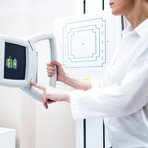
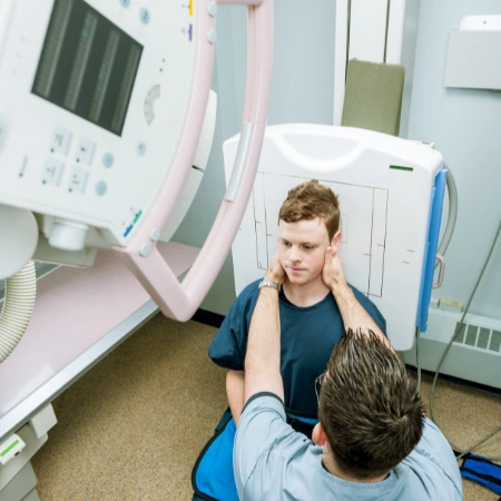
Imaging the Lower Extremity
An interactive CE course in Imaging of the Lower Extremity.
- Approved by the ASRT (American Society of Radiologic Technologists) for 2.75 CE Credits
- Subscription duration: 365 days from purchase date
- Voiceover available
- Downloadable transcript available
- *NEW* Video format available with subtitles
- Meets the CE requirements of the following states: California, Texas, Florida, Kentucky, Massachusetts, and New Mexico
- Meets ARRT® CE reporting requirements
- Accepted by the NMTCB®
- Hassle-free 30-day full refund policy*
X-ray were discovered on November 8, 1895, by Wilhelm Conrad Roentgen. Almost immediately these new ‘rays’ were recognized for their amazing ability to penetrate the human body and demonstrate the skeletal system and other internal structures. From its primitive beginning, diagnostic imaging has undergone numerous changes in the methods of imaging, the methods of developing, method of image display, and the methods of storing the image. However, there has been little change in the patient positioning aspect of diagnostic radiography.
Regardless of the technology used, proper patient positioning is critical. These modules will review important imaging aspects including effective communication skills, the importance of standard precaution, ALARA, pre and post patient instructions and the essential image positioning and image critique skills needed to create quality radiographs. Module 1 reviews imaging of the hips though knees. Module 2 reviews imaging the lower leg to toes.
| Discipline | Major content category & subcategories | CE Credits provided |
| RAD-2017 | Patient Care | |
| Patient Interactions and Management | 0.50 | |
| RAD-2017 | Safety | |
| Radiation Protection | 0.25 | |
| RAD-2017 | Procedures | |
| Extremity Procedures | 2.00 | |
| RA-2018 | Patient Care | |
| Patient Management | 0.25 | |
| RA-2018 | Safety | |
| Patient Safety, Radiation Protection, and Equipment Operation | 0.25 | |
| RA-2018 | Procedures | |
| Musculoskeletal and Endocrine Sections | 0.50 | |
| RAD-2022 | Patient Care | |
| Patient Interactions and Management | 0.50 | |
| RAD-2022 | Safety | |
| Radiation Protection | 0.25 | |
| RAD-2022 | Procedures | |
| Extremity Procedures | 2.00 | |
| RA-2023 | Patient Care | |
| Patient Management | 0.25 | |
| RA-2023 | Safety | |
| Patient Safety, Radiation Protection, and Equipment Operation | 0.25 | |
| RA-2023 | Procedures | |
| Musculoskeletal and Endocrine Sections | 0.50 |
Section 1: Hips through Knees
- Patient Care
- Technologist set-up
- Hip positioning techniques
- Femur positioning techniques
- Knee positioning techniques
- Patella positioning techniques
Section 2: Lower Legs to Toes
- Tibia/Fibula positioning techniques
- Ankle positioning techniques
- Calcaneus positioning techniques
- Foot positioning techniques
- Toes positioning techniques
|
Get it now!
One-time payment. No hidden fees. No extra charges per credit.
|
|
|
Unlimited CE Credits
For Only $49.99
1 – year Access – Doesn’t Auto-Renew
Guaranteed 30-day Refund Policy