
Sports Injury Imaging in Soccer: World Cup Edition!






- Introduction
- Anterior Cruciate Ligament (ACL) Injuries
- Hamstring Muscle Strains
- Meniscal Tears
- Ankle Ligament (ATFL) Sprains
- Stress Fractures & Bone Edema
- Quick Reference: Imaging Protocols for Soccer Injuries
- Conclusion
Introduction
Soccer (or football, as it’s known worldwide) is a high‑intensity sport with rapid sprints, sudden pivots, jumps, and frequent contact, all of which increase the risk of injuries. Imaging plays a crucial role not just in diagnosis, but also in planning treatment and guiding safe return to play.
In the spirit of the World Cup 2026, this article explores common soccer injuries and highlights the imaging protocols used to evaluate them, giving technologists a practical, “on-the-pitch” perspective.
Anterior Cruciate Ligament (ACL) Injuries
The anterior cruciate ligament (ACL) is one of the key stabilizers of the knee, preventing excessive forward movement and rotation of the tibia. In soccer, ACL injuries most often occur during non‑contact events, like sudden deceleration, pivoting, or awkward landings after a jump, but can also happen through collisions. For players, an ACL tear is not only painful but can sideline them for months, making timely and accurate imaging critical.
Imaging is essential for confirming the diagnosis, assessing the tear pattern, checking for associated meniscus or cartilage injuries, and planning rehabilitation or surgical reconstruction.
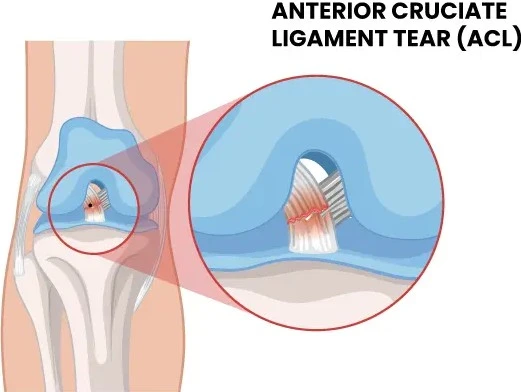
Best imaging protocols for an ACL injury:
-
MRI (gold standard)
-
Sequences: Multiplanar PD-weighted and fat-suppressed (FS) sequences in sagittal, coronal, and axial planes
-
Optional: 3D isotropic sequences on 3T scanners for pre-surgical planning
-
Purpose: Evaluate ligament integrity, bone bruises, associated meniscus tears, cartilage status
-
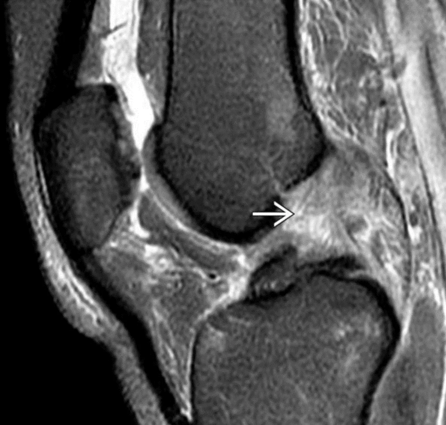 Sagittal PD‑weighted fat‑suppressed (PDWI FS) MRI of the knee showing an acute complete tear of the anterior cruciate ligament (ACL)
The image here is a Sagittal PD‑weighted fat‑suppressed (PDWI FS) MRI of the knee showing an acute complete tear of the anterior cruciate ligament (ACL) (white solid arrow). The ligament fibers are absent in the proximal half, and there is extensive edema in the proximal intercondylar notch. The arrow points to where the ACL should normally be. Clinically, patients often report a loud “pop” at the time of injury, severe pain, rapid swelling, knee instability, and difficulty bearing weight or moving the joint. This image highlights the classic appearance of an acute ACL tear, emphasizing the importance of MRI in confirming the diagnosis and guiding treatment.
-
Radiographs (initial evaluation): AP, lateral, and optional tunnel view to exclude avulsion or fracture.
-
Ultrasound: Evaluate surrounding soft tissue; limited for ACL itself
Famous Player Cases: Neymar Jr.
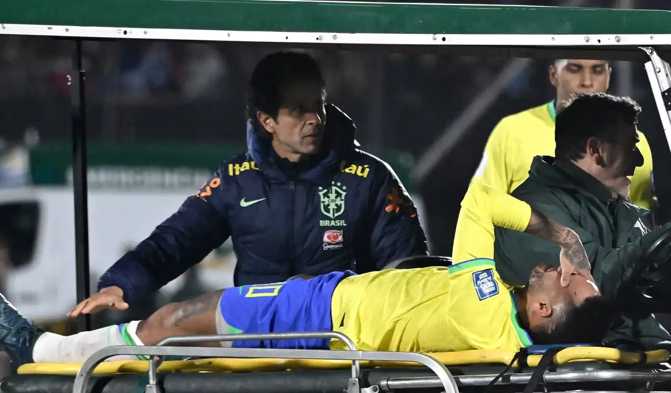
A famous recent example was Neymar, who suffered a ruptured ACL and meniscus in his left knee during a 2026 FIFA World Cup qualifying match against Uruguay, when he was taken off on a stretcher and later required surgical reconstruction after imaging confirmed the injury. The diagnosis meant an extended period away from play and an intense rehabilitation process before he was able to return to competitive action
Hamstring Muscle Strains
Hamstring muscle strains are among the most common injuries in soccer because the hamstrings are under tremendous stress during high-speed running, explosive acceleration, and sudden deceleration, all actions that define the modern game. They occur when one or more of the hamstring muscles (semimembranosus, semitendinosus, and biceps femoris) are overstretched or overloaded beyond their capacity, leading to tearing of muscle fibers (Grades 1-3 depending on severity).
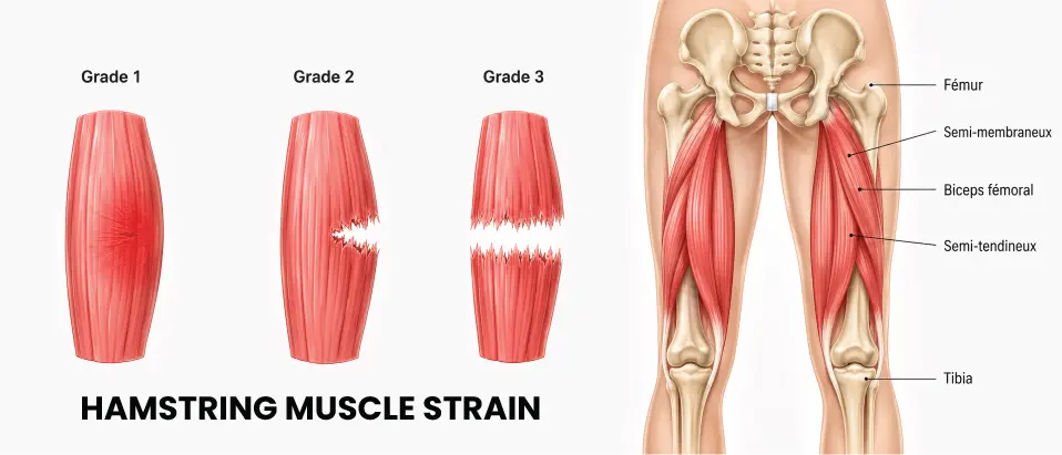
Imaging has a key role in determining the extent and location of the injury, guiding treatment and influencing how quickly a player may return to competition. It also helps differentiate a simple strain from a more significant tear or avulsion, which may require more targeted rehabilitation strategies.
Best imaging protocols for a hamstring muscle strain injury:
-
MRI (Gold Standard):
-
Ideal for visualizing muscle fiber disruption, hematoma, and edema on fluid-sensitive sequences such as STIR or T2-weighted fat-suppressed images.
-
Helps classify the severity of the strain and detect injuries near the myotendinous junction.
-
Can also assess associated injuries in adjacent soft tissues.
-
Sequences:Axial and coronal T2-weighted FS, STIR sequences for edema
-
Purpose:Assess muscle fiber disruption, hematoma, myotendinous junction integrity, and edema pattern
-
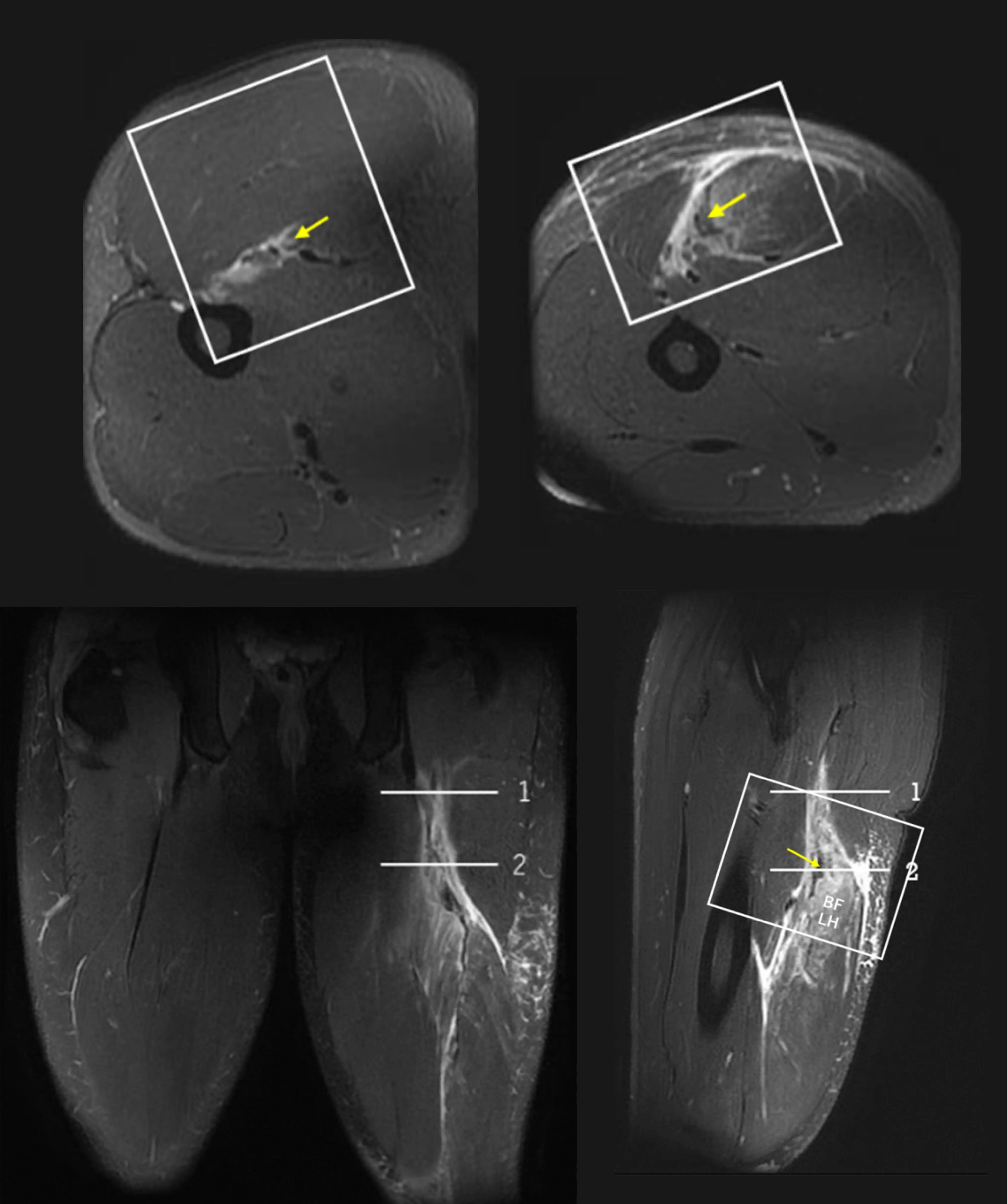 MRI of a hamstring tear. Multi-planar T2-weighted fat-suppressed images show fiber rupture, distal displacement, and hematomas along the semitendinosus and biceps femoris.
Ultrasound:
-
Useful for dynamic assessment, especially in partial tears, and can be used to monitor healing over time.
-
Real-time imaging allows evaluation during muscle activation and comparison with the contralateral limb.
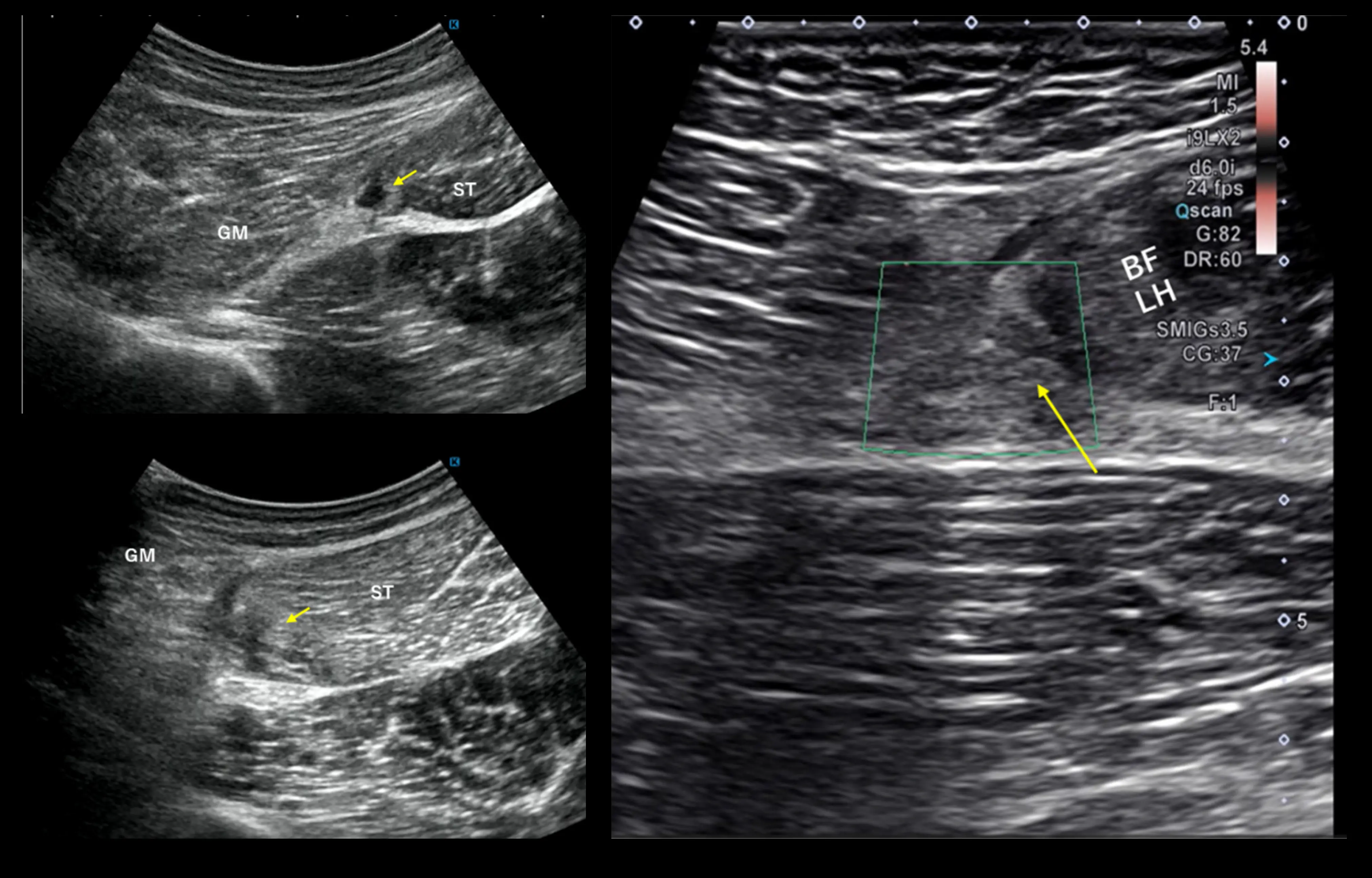 Ultrasound of a hamstring tear, useful for dynamic assessment of partial tears and monitoring healing over time.
Players with hamstring strains often describe a sudden, sharp pain in the back of the thigh, frequently accompanied by immediate weakness, followed by swelling and a temporary inability to continue play. MRI may show focal muscle edema and, in more severe cases, clear disruption of muscle fibers. Properly documented imaging drives clinical decisions about load progression, physical therapy, and return-to-play timing.
Famous Player Cases: Lamine Yamal
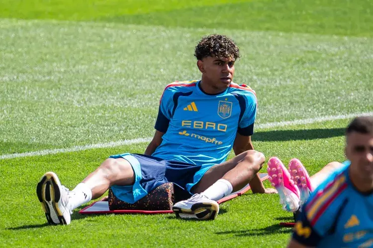
In elite soccer, hamstring injuries are so frequent that teams often lose valuable training time and matches due to time spent rehabilitating these strains. A high-profile example in the lead-up to the 2026 FIFA World Cup is Lamine Yamal, who missed the end of the club season with a hamstring injury and was managed cautiously by Barcelona and Spain’s medical staff as the tournament kicked off.
Famous Player Cases: Raphinha
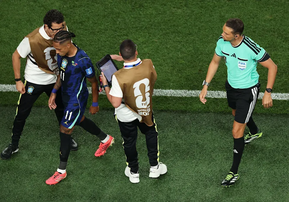
Another high-profile hamstring injury emerged during the 2026 FIFA World Cup, involving Brazil’s Raphinha. In a Group C matchup against Haiti, the Barcelona winger felt a sudden, sharp pain in the back of his right thigh during an explosive sprint, forcing an early substitution before halftime. The Brazilian Football Confederation (CBF) medical staff prioritized immediate pitchside video review to examine the biomechanics of the acceleration, followed by an urgent MRI scan. The imaging protocol revealed a Grade 1 hamstring strain located in his right thigh.
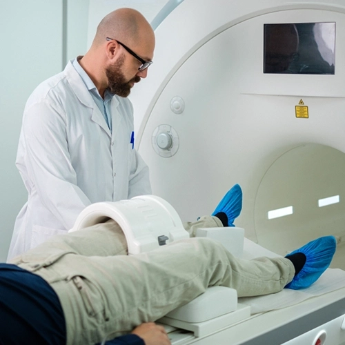
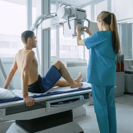
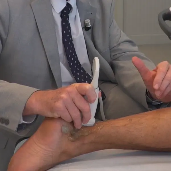
Advance your musculoskeletal imaging skills today!
Explore our hands-on MRI, X-ray and ultrasound courses designed to elevate your clinical expertise and confidence.
Meniscal Tears
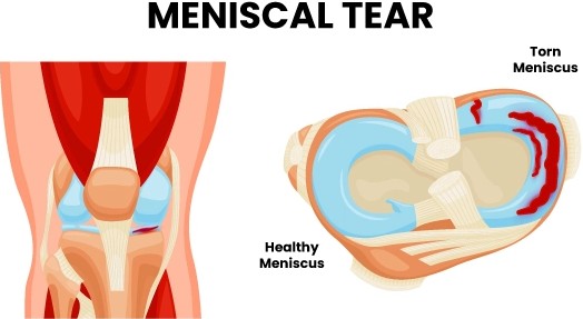
Meniscal tears are injuries to the C-shaped cartilage pads (medial and lateral menisci) that sit between the femur and tibia, acting as shock absorbers and stabilizers for the knee joint. In soccer, meniscal tears often occur from twisting motions, sudden pivots, or in conjunction with other injuries like ACL tears, especially when a player changes direction rapidly or lands awkwardly. These tears can be radially, horizontally, bucket-handle, or flap patterns, and the exact morphology affects both management and prognosis.
Because meniscal tears are often subtle on physical exam and may mimic other internal derangements, high-quality imaging is essential. MRI is the primary modality used to confirm the tear, characterize its type, and inform surgical vs. conservative management, making the role of the rad tech crucial in acquiring diagnostic sequences.
Best imaging protocols for meniscal tear injuries:
-
MRI (Gold Standard):
-
Sequences:Sagittal and coronal PD-weighted + fat-suppressed views in sagittal, coronal, and axial planes provide optimal visualization of meniscal morphology and tear patterns.
-
Fluid-sensitive sequences help demonstrate displaced fragments (e.g., bucket-handle tears).
-
Additional sequences may assess associated injuries such as ligamentous tears or bone bruises.
-
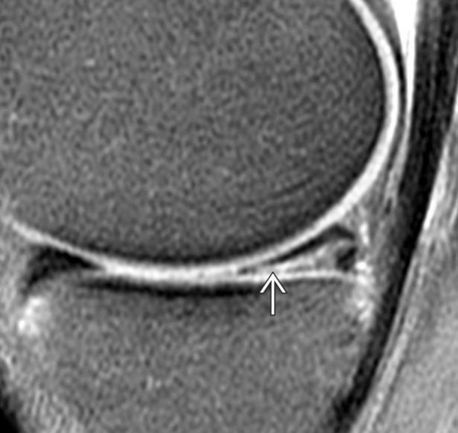
Sagittal PD-weighted fat-suppressed MRI showing a horizontal cleavage tear of the posterior horn of the medial meniscus (white arrow). The tear extends to the inferior articular surface, while the anterior horn remains intact. This image highlights the classic appearance of a meniscal tear and aids in diagnosis and treatment planning.
Famous Player Cases: Gavi
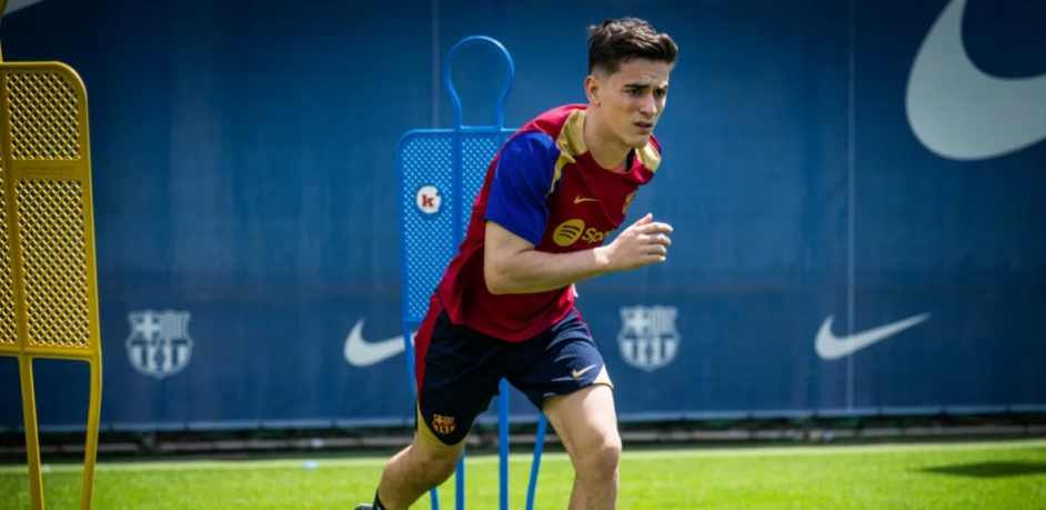
Meniscal tears are not only common, they also affect some of the sport’s biggest stars. For example, Gavi suffered a meniscal injury during the 2025-26 season, which was also confirmed with MRI and required surgical management, resulting in several weeks away from play as part of his rehabilitation. Such high-profile cases underline the importance of clear, high-quality imaging to accurately diagnose the extent and pattern of a meniscal tear, critical information that guides treatment decisions and return-to-play timing for elite athletes.
Ankle Ligament (ATFL) Sprains
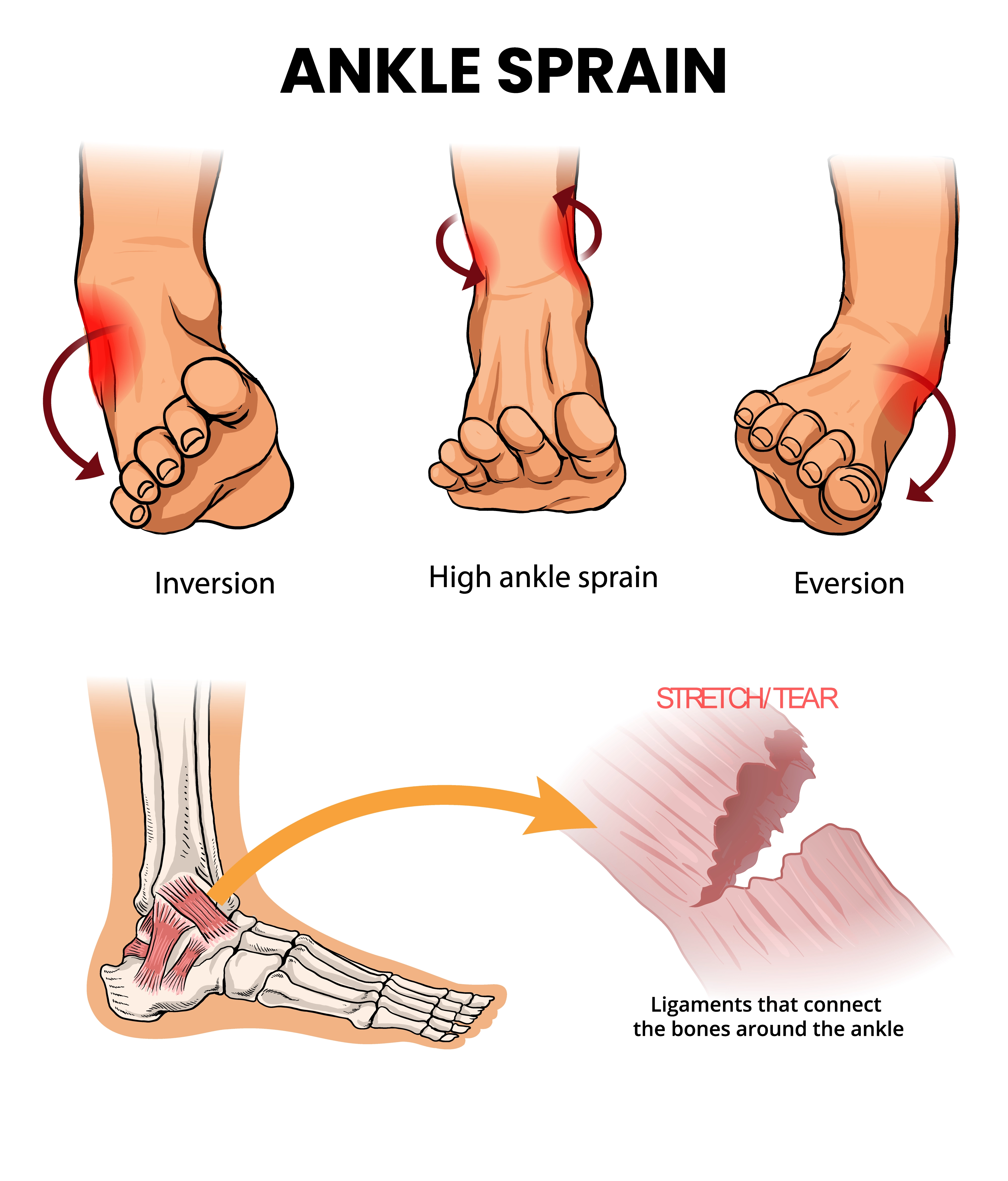
One of the most common acute injuries in soccer is a sprain of the lateral ankle ligaments, especially the anterior talofibular ligament (ATFL). These injuries typically occur when a player’s foot lands awkwardly after a jump, collides with another player, or suddenly inverts (rolls inward), placing excessive strain on the ligaments that stabilize the outside of the ankle. Although often thought of as “just a sprain,” a severe ATFL tear can lead to prolonged symptoms, instability, and time off the pitch.
Because the ankle joint is small but complex, imaging plays an essential role in determining the severity of the ligament injury, identifying associated fractures or bone bruises, and guiding treatment decisions, from conservative management with physical therapy to possible surgical repair in high-grade tears or chronic instability.
Best imaging protocols for ATFL sprains:
-
Radiographs
-
AP, lateral, and mortise views
-
First-line in acute ankle trauma.
-
Excellent for ruling out fractures or avulsion injuries at ligament attachment sites.
-
Quick and widely available in most imaging departments.
-
-
Ultrasound
-
Dynamic evaluation of ligament integrity during stress maneuvers using a high-frequency linear probe, dynamic stress views for ligament laxity or rupture.
-
Shows partial vs. complete tears and associated soft tissue swelling in real time.
-
Highly useful in clinics or emergency settings as a complementary tool.
-
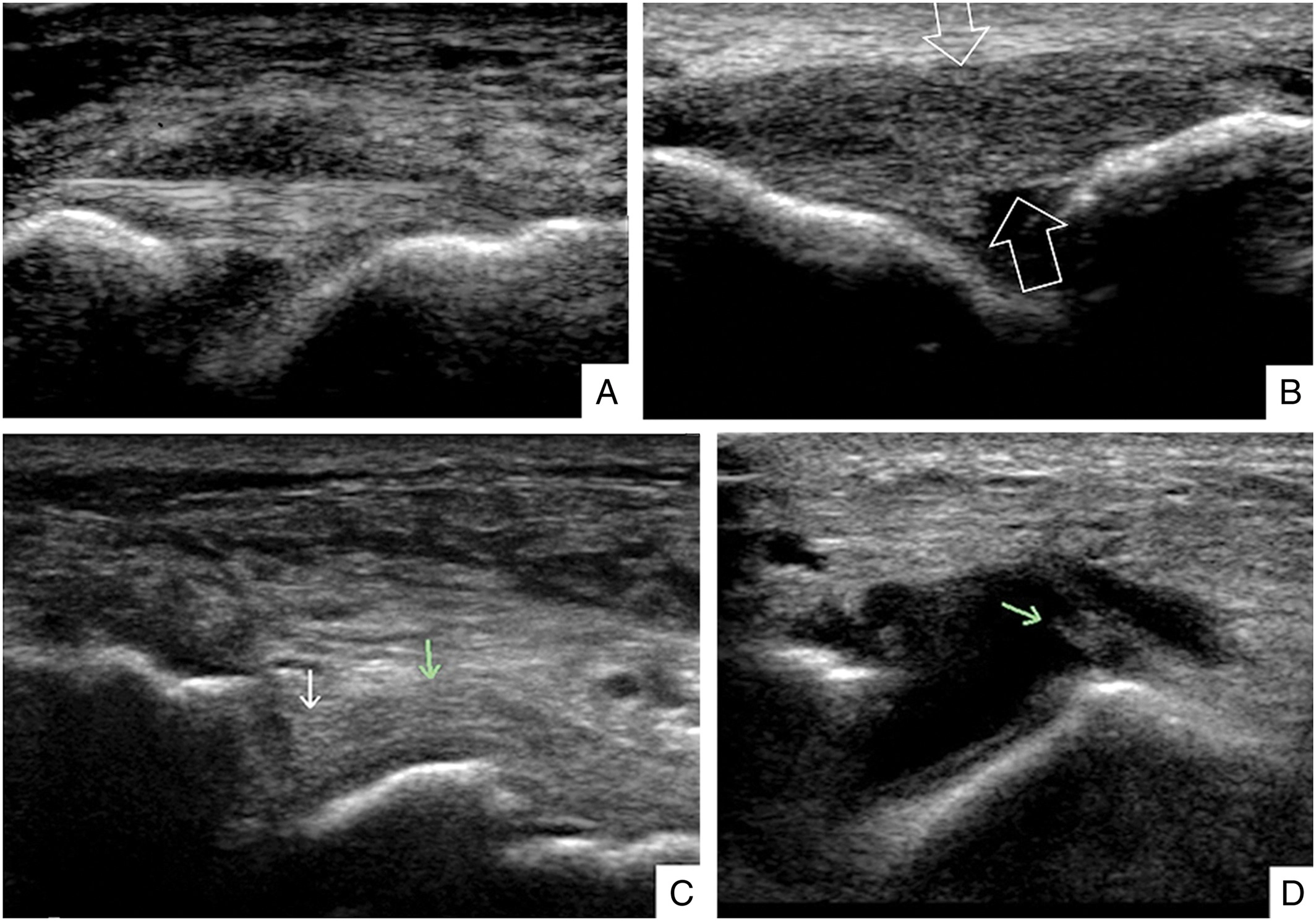
Ultrasound images demonstrating anterior talofibular ligament (ATFL) injuries of varying severity. (A) Grade I, ligament fibers remain intact with superficial edema. (B) Grade II, ligament intact but lax and oedematous. (C and D) Grade III, complete rupture of the ATFL, accompanied by joint effusion. Ultrasound allows dynamic, real-time assessment of ligament integrity and associated soft tissue changes, making it invaluable for grading ankle sprains, guiding treatment decisions, and monitoring recovery.
-
MRI
-
Performed when symptoms persist, when there’s clinical suspicion of complex multi-ligament injuries, or when bone/chondral injuries are suspected.
-
Sequences:T1, T2 FS, and proton density FS sequences to assess ligament fibers, effusion, and associated bone marrow edema.
-
Provides clear delineation of ligament fibers, surrounding soft tissues, and secondary findings like bone edema.
-
Famous Player Cases: Chris Richards
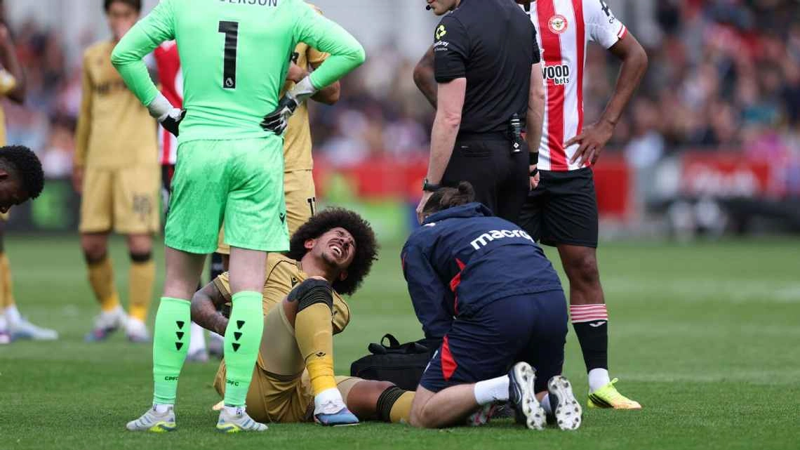
A notable recent ankle ligament injury involved Chris Richards, defender for Crystal Palace and the U.S. Men’s National Team, who suffered tear(s) of ligaments in his ankle during a Premier League match in May 2026. Imaging was used to assess the injury and guide his ongoing recovery, and the injury raised questions about his fitness ahead of the 2026 FIFA World Cup as he underwent rehabilitation and monitored return-to-play progress.
Stress Fractures & Bone Edema
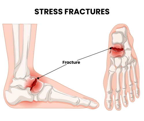
Soccer’s demands high mileage, frequent accelerations and decelerations, and an unpredictable playing surface can subject bones to repetitive loading that sometimes leads to microscopic damage within the bone itself. This process begins with bone marrow edema, a response to abnormal stress, and if the overload continues without adequate recovery, it can progress to a stress fracture. In soccer players, these injuries most commonly affect the tibia and metatarsals, where concentrated forces occur during running and jumping.
Early recognition is critical: if caught in the bone edema stage, athletes can modify activity and prevent full cortical fracture; if not, a complete stress fracture can sideline a player for weeks or even months.
Best imaging protocols for stress fractures and bone edema:
-
MRI (Highly Sensitive for Early Injury):
-
The modality of choice in the acute setting.
-
Shows marrow edema before changes are visible on X-rays.
-
Sequences:Coronal and sagittal STIR or T2 FS; optional axial PD FS (fluid-sensitive sequences) will demonstrate diffuse high signal in the affected bone.
-
Helpful for differentiating bone stress injury from other causes of pain such as muscle strain or contusion.
-
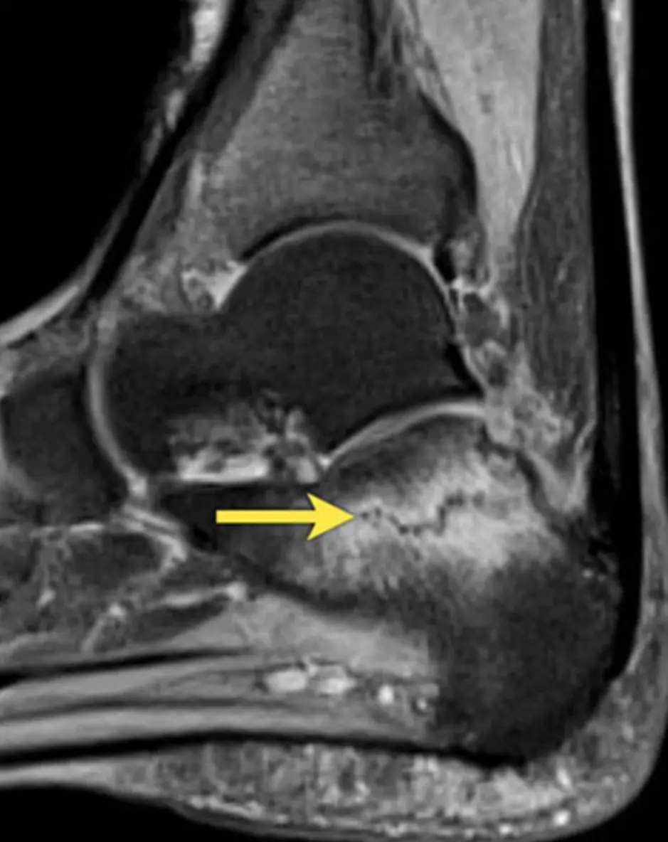
MRI of a stress fracture in the calcaneus (yellow arrow) showing associated bone marrow edema. This type of overuse injury is common in runners and athletes, and MRI is essential for early detection and management.
-
Radiographs:
-
Often normal early on, especially in the first 2-3 weeks.
-
AP, lateral views; may be normal early in the injury.
-
With time, the follow-up radiographs may show periosteal reaction or a visible fracture line as healing begins.
-
Still a valuable initial test since it’s widely available, quick, and inexpensive.
-
Famous Player Cases: Jack Grealish
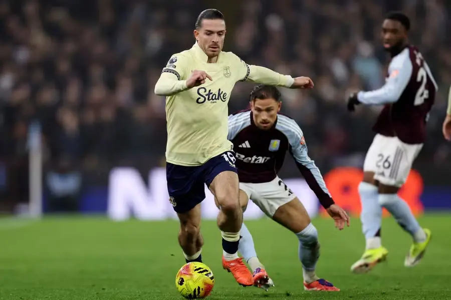
One recent stress fracture case involved Jack Grealish, who in early 2026 suffered a foot stress fracture confirmed by imaging, underwent surgery, and saw his World Cup aspirations disrupted as a result, a reminder of how overuse bone injuries can impact even elite players when not identified and managed promptly.
Quick Reference: Imaging Protocols for Soccer Injuries
The table below provides a quick reference summary of optimal imaging protocols for the most common soccer injuries we’ve discussed, highlighting each modality’s key benefits for rad techs in clinical practice.
| Injury Type | First-Line Imaging | Optimal / Detailed Imaging Protocol |
|---|---|---|
| ACL Tear | X-ray: AP, lateral (exclude fracture/avulsion) | MRI (gold standard): Multiplanar PD-weighted + fat-suppressed sequences in sagittal, coronal, axial planes; optional 3D isotropic sequences on 3T; assesses ligament integrity, bone bruises, meniscus, cartilage |
| Hamstring Strain | Clinical exam + Ultrasound (dynamic, partial tear assessment) | MRI: Axial & coronal T2 FS, STIR sequences for edema; evaluates muscle fiber disruption, hematoma, myotendinous junction involvement; monitor healing over time |
| Meniscus Tear | MRI knee (standard PD FS sequences) | MRI Detailed: Sagittal & coronal PD FS sequences; optional axial plane; identifies tear type (bucket-handle, horizontal, vertical), fragment displacement, and associated cartilage/ligament injury |
| Ankle Sprain (ATFL) | X-ray: AP, lateral, mortise views | Ultrasound: High-frequency linear probe with dynamic stress views; MRI: T1, T2 FS, PD FS sequences to assess ligament fibers, effusion, and bone marrow edema; useful for high-grade or multi-ligament injuries |
| Stress Fracture / Bone Edema | X-ray: AP, lateral (often normal early) | MRI: Coronal & sagittal STIR or T2 FS; optional axial PD FS; detects early marrow edema, differentiates from soft tissue injury; follow-up MRI or CT if fracture progression suspected |
Note:Routine radiographs are often performed first in acute injuries to exclude fractures and alignment issues, but MRI remains the modality of choice for soft tissue and early stress injuries.
Conclusion
Soccer injuries cover a range from soft tissue strains to severe ligamentous ruptures. Imaging selection must be guided by injury mechanism, expected pathology, and clinical urgency. Soccer’s global profile and the World Cup spotlight offer opportunities to educate clinicians and radiologists on optimized imaging protocols and the utility of advanced modalities like MRI and ultrasound for precise diagnosis.
References
-
Neymar ACL injury– FIFA.Neymar sustains injury in Brazil vs Uruguay qualifier.
https://www.fifa.com/en/news -
Hamstring strain imaging– MDPI.Ultrasound Diagnosis of Hamstring Muscle Complex Injuries Focus on Originate Tendon Structure – Male University Rugby Players.
https://www.mdpi.com -
Lamine Yamal hamstring injury– BarcaBlaugranes.Barcelona clear Lamine Yamal to play for Spain at World Cup 2026 but set conditions after hamstring injury.
https://www.barcablaugranes.com/ -
Raphinha hamstring injury– sports.yahooBrazil star Raphinha leaves World Cup match against Haiti with injury.
https://sports.yahoo.com/ -
Gavi meniscal tear– Reuters.Barca’s Gavi undergoes knee surgery for meniscus injury.
https://www.reuters.com/sports/soccer/ -
ATFL sprain imaging– Wiley Online Library.Ultrasound assessment of lateral ankle ligament injuries.
https://onlinelibrary.wiley.com/ -
Jack Grealish foot stress fracture (2026)– Flashscore.Everton’s Jack Grealish to miss remainder of season after foot surgery.
https://www.flashscore.com/news/ -
Stress fracture / bone edema imaging– Radiology Assistant.MRI Examination – Musculoskeletal Imaging.
https://radiologyassistant.nl -
Chris Richards ankle ligament injury (2026)– ESPN.Chris Richards suffers ankle ligament injury ahead of 2026 World Cup.
https://www.espn.co.uk/football